Our group studies biomineralization in various biological systems. Two of these studies have been published in the prestigious journal "Science".
The crystallographic structure of vaterite, the least stable polymorph of calcium carbonate, has been a long subject of debate among scientists. A research done by our group on the spicules of Herdmania momus has discovered that vaterite is in fact composed of two different crystallographic structures, a major structure with hexagonal symmetry and another structure with yet unknown symmetry that coexists as nanodomains within the major structure. The coexistence of these two structures was found by means of High-Resolution Transmission Electron Microscopy. These findings helped revealing the crystallographic structure of vaterite that has been a puzzle to scientists for over a century.
The brittle star Ophiocoma wendtii is able to detect predators and escape from them. This feature was remarkably puzzling to researchers since no visualization system was known in this specie. It was discovered in 2001 that this brittle star has a unique visualization system composed of an array of single crystal calcitic microlenses positioned on his armplates.
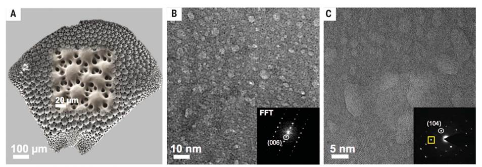
Figure 1. Electron microscopy. (A) HRSEM image showing an entire DAP and a higher magnification insert. (B) HRTEM in bright-field mode revealing brighter nanodomains, although the FFT pattern is that of a single crystal. Higher magnification (C) of an area in (B) shows a lattice image undisrupted by the nanodomains which demonstrate coherent interfaces with the lattice. The insert shows an electron diffraction image from this area.
These microlenses focus the light onto bundles of nerves located below them. In addition, their axis is directed parallel to the lens' optical axis to avoid birefringence. A research done by our group has found a unique and novel strategy for strengthening and toughening the microlenses of the brittle star Ophiocoma Wendtii. The above is achieved by the presence of Mg- rich nanoparticles that are coherent with the calcitic matrix. The presence of these nanoparticles induces compressive stresses into the matrix thus causing the whole structure to be pre-compressed. These findings were achieved using state- of- the- art techniques such as High-Resolution Transmission Electron Microscopy and High-Resolution Powder XRD using synchrotron radiation.
We further established that the presence of high-Mg nanoparticles embedded within lower-Mg calcite matrices is a widespread strategy utilized by various organisms from different kingdoms and phyla to improve the mechanical properties of their high-Mg calcite skeletons. We show that such phase separation and the formation of high-Mg nanoparticles are most probably achieved through spinodal decomposition of an amorphous Mg-calcite precursor. Such decomposition is independent of the biological characteristics of the studied organisms belonging to different phyla and even kingdoms but rather, originates from their similar chemical composition and a specific Mg content within their skeletons, which generally ranges from 14 to 48 mol % of Mg. We show evidence of high-Mg calcite nanoparticles in the cases of six biologically different organisms all demonstrating more than 14 mol % Mg-calcite and consider it likely that this phenomenon is immeasurably more prevalent in nature. We also establish the absence of these high-Mg nanoparticles in organisms whose Mg content is lower than 14 mol %, providing further evidence that whether or not spinodal decomposition of an amorphous Mg-calcite precursor takes place is determined by the amount of Mg it contains.
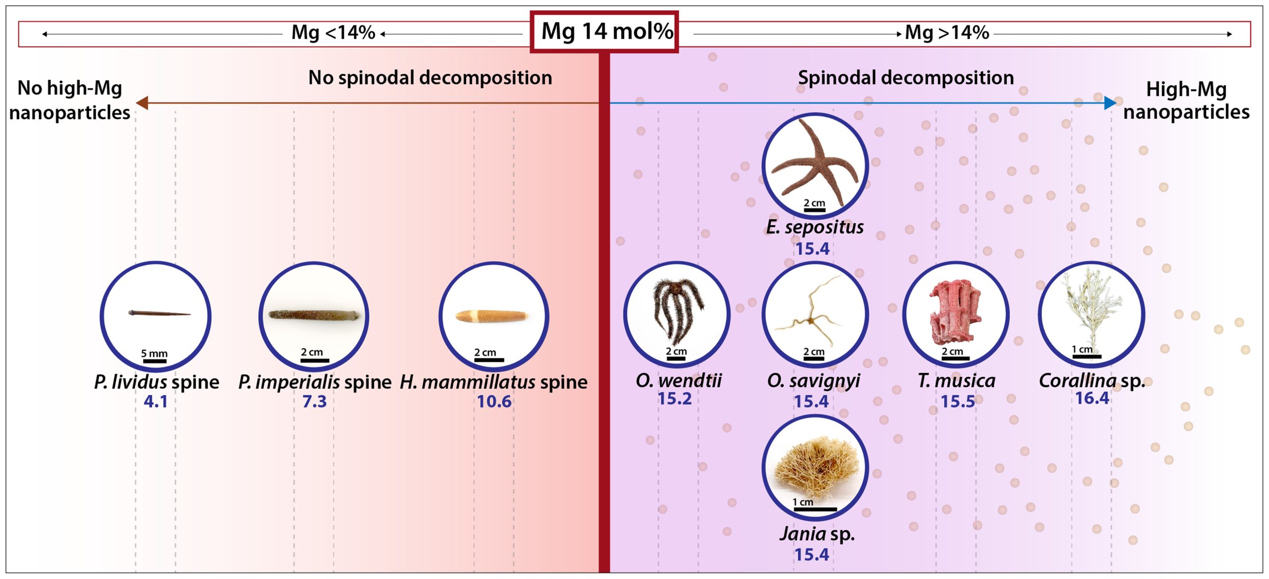
The various studied biomineralized tissues divided into two categories based on their Mg content. High-Mg nanoparticles are present only for those with an average Mg content higher than 14 mol %.
The valuable knowledge gained from this biostrategy significantly impacts the understanding of how biominerals, although composed of intrinsically brittle materials, can effectively resist fracture. Moreover, our theoretical calculations clearly suggest that the formation of Mg-rich nanoprecipitates greatly enhances the hardness of the biomineralized tissue as well.
Publications:
Bianco-Stein N, Polishchuk I, Lang A, Portal L, Dejoie C, Katsman A and Pokroy B*. High-Mg calcite Nanoparticles within a Low-Mg Calcite Matrix: a Widespread Phenomenon in Biomineralization. PNAS 2022, 119(16):e21201771199.
Bianco-Stein N, Polishchuk I, Lang A, Atiya G, Villanova J, Zaslansky P, Katsman A and Pokroy B*. Structural and Chemical Variations in the Calcitic Segments of Coralline Red Algae Lead to Improved Crack Resistance. Acta Biomater 2021; 130:362.
Bianco‐Stein N, Polishchuk I, Seiden G, Villanova J, Rack A, Zaslansky P and Pokroy B. Helical Microstructures of the Mineralized Coralline Red Algae Determine Their Mechanical Properties. Adv Sci 2020; 7:2000108.
Polishchuk I, Aronhime Bracha A, Bloch L, Levy D, Kozachkevich S, Etinger-Geller Y, Kauffmann Y, Burghammer M, Giacobbe C, Villanova J, Hendler G, Sun C Y, Giuffre A.J, Marcus M.A, Kundanati L, Zaslansky P, Pugno N.M, Gilbert Pupa U. P. A., Alex Katsman A and Pokroy B. Coherently aligned nanoparticles within a biogenic single crystal: A biological prestressing strategy. Science 2017; 358:1294. Click here for FREE download.
Pokroy B, Fieramosca JS, Von Dreele RB, Fitch AN, Caspi EN, Zolotoyabko E. Atomic structure of biogenic aragonite. Chem Mater 2007; 19:3244.
Kabalah-Amitai L, Mayzel B, Kauffmann Y, Fitch AN, Bloch L, Gilbert PUPA, Pokroy B. Vaterite Crystals Contain Two Interspersed Crystal Structures. Science 2013; 340:454.
Pokroy B, Fitch AN, Marin F, Kapon M, Adir N, Zolotoyabko E. Anisotropic lattice distortions in biogenic calcite induced by intra-crystalline organic molecules. J Struct Biol 2006; 155:96.